World's most detailed scans will reveal how brain works
- Published

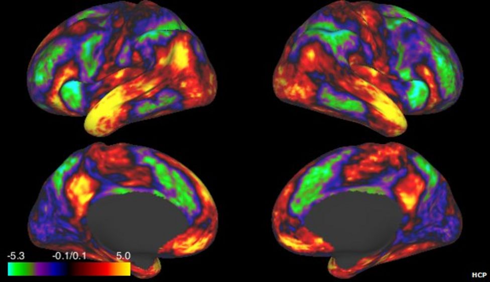
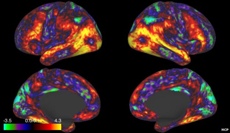

Scientists say they have published the most detailed brain scans "the world has ever seen" as part of a project to understand how the organ works.
The aim of the project is to determine how a person's brain structure influences their talents and behaviour.
Researchers involved in the so called Human Connectome Project have published the scans of 68 adults in the study.
They eventually hope to scan 1,200 people and also collect details of their behavioural traits and DNA.
The information is made freely available to neuroscientists in their quest to unlock the secrets of the human brain.
The project leader, Prof David Van Essen of Washington University in St Louis, told BBC News that sharing the data with the international community of researchers would spur rapid advances in brain science.
"We are very optimistic that as the community delves in and begins working on these data sets, they will reveal new insights into the brain circuits of healthy adults," he said.
Subjects involved in the project have their brain scanned for a total of four hours. For part of that time, they carry out a battery of tasks, which include arithmetic, listening to stories, gambling and moving parts of their body.
Volunteers also engage in tests that assess their skills and abilities. In addition, DNA samples are taken.
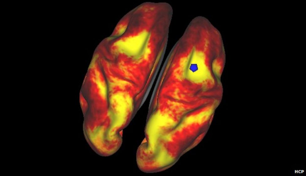
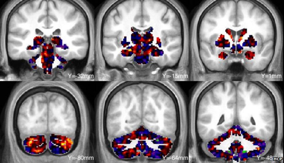
The scans are essentially a wiring diagram for each person's brain.
They show how different parts are connected by nerve fibres and also the thickness of the bundles, which is thought to be an indication of the importance or strength of a particular connection- a so-called "structural map".
Scanning can also show which parts of the brain are activated for particular tasks - known as a "functional map".
With all this information, researchers will be able to see if an individual's brain wiring is related to their skills, such as musicality, sociability and aptitude for science or maths.
Neural circuitry
According to Oxford University's Dr Tim Behrens, who is collaborating with Prof Van Essen, the study will "uncover which neural pathways are important in determining human behaviours".
The eventual aim of the project is to understand how the healthy human brain is wired and how differences between individuals make each person unique - shaping their personalities and their capacity to think and feel.
Prof Van Essen is excited by what may be revealed.
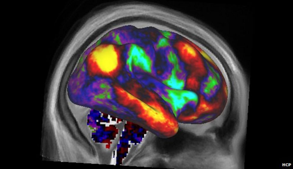
The BBC's Pallab Ghosh gets a look at how his brain is wired
"We have the highest quality data of the entire human brain that the world has ever seen. The question is that with more cutting edge (scanning) methods, how much can we decipher the circuits that give us our distinctive capabilities?" he said.
By learning more about how the healthy human brain works, the research will inevitably be of use to those studying brain disorders, such as Alzheimer's and Parkinson's.
Among those who will be delving through the data is Ed Bullmore, a professor of psychiatry at Cambridge University. He believes that psychiatric problems, such as schizophrenia, drug addiction and obsessive compulsive disorder generally arise from irregular brain development.
Dementia
"We'll have a better opportunity to understand these disorders once we have a better grip on normal brain development", he told BBC News.
The research data is also likely to help those seeking to stem or slow down dementia. The study will undoubtedly lead to better ways of identifying those most at risk from their brain scans.
An important aim of the £26m ($40m), five-year, US-government-funded project is to share the data with scientists across the world.
Those behind the project were inspired by the way that the sharing of information gleaned by the Human Genome project has spurred the acceleration of genetic science. But this concept has been lower to take hold in brain imaging, and the associated emerging field of neuroinformatics.
The problem has been the sheer complexity of the data and the ensuing processing and analysis of the information.
For example, the images just released of the 68 subjects take up about two terabytes of computer memory, which is two thousand billion bytes, enough to fill several hundred DVDs.
The Human Connectome Project has therefore developed a database called ConnectomeDB to make sharing of brain images much easier.
"In my optimistic view, I believe this will spur an acceleration in neuroinformatics which will be able to acquire and analyse data [from brain scans] in more powerful ways than has been possible up to this point.
"This in turn could lead to a transformative set of developments that could accelerate our understanding of the brain," Prof Van Essen told BBC News.
Follow Pallab on Twitter
- Published17 February 2013
- Published7 February 2013